Blood
Diagram
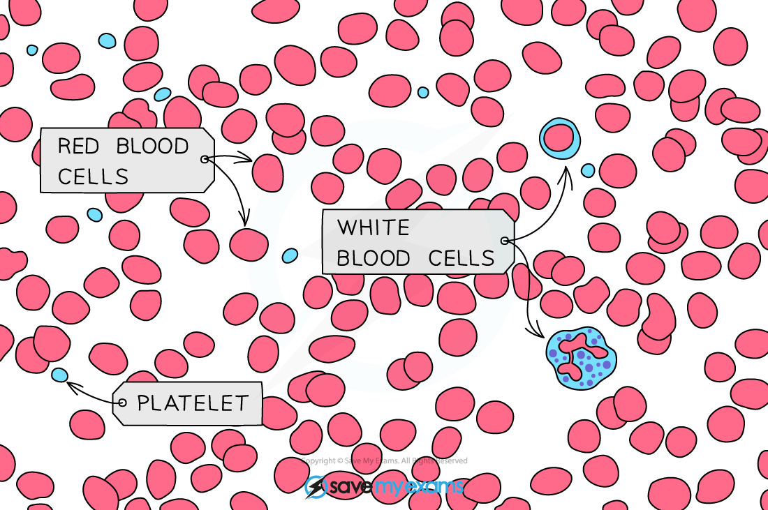
Components as viewed in diagrams
- Red blood cells: Very numerous. Circular discs with a lighter center (biconcave shape). Contains no nucleus.
- White blood cells: Larger than red blood cells, fewer in numbers. Has a dark-staining nucleus.
- Phagocytes: Easily identified by their lobed (segmented) nucleus and an irregular, flexible outer cell membrane.
- Lymphocytes: Identified by a large, round nucleus that occupies nearly the entire volume of the cell, leaving only a thin rim of cytoplasm inside.
- Platelets: Irregular cell fragments. Appear as small dots scattered between the red blood cells.
- Plasma: Empty space surrounding the blood cells.
Functions of blood components
| Component | Functions |
|---|---|
| Red blood cells | Oxygen transport from lungs to respiring tissues. They contain hemoglobin, an iron-rich protein that binds reversibly to oxygen to form oxyhemoglobin. |
| Phagocytes | Protect the body by engulfing and digesting pathogens (bacteria/viruses) via a process called phagocytosis. |
| Lymphocytes | Protect the body by producing antibodies. These proteins bind to antigens on pathogens, clumping them together or marking them for destruction by phagocytes. |
| Platelets | Responsible for initiating the blood clotting process when a blood vessel is damaged. |
| Plasma | Liquid that serves as the transport medium for blood cells, ions, nutrients (glucose, amino acids), waste products (urea and carbon dioxide) and hormones. |
Roles of blood clotting
- Prevents excessive blood loss by sealing the broken vessel.
- Prevents the entry of pathogens into the bloodstream, which could otherwise cause infections.
Mechanism
- Activating platelets: Damage to the blood vessel lining exposes collagen fibers, causing platelets to adhere to the site and release clotting factors.
- Protein conversion: These factors trigger a process that converts a soluble plasma protein called fibrinogen into an insoluble fibrous protein called fibrin.
- Mesh formation: The fibrin strands stretch across the damaged area, forming a sticky, microscopic mesh.
- Trapping cells: Red blood cells and additional platelets flowing past become trapped within the fibrin mesh, forming a solid plug (clot) that later hardens into a scab.