Blood Vessels
Diagram
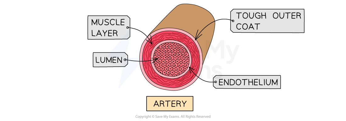
Structure of arteries, veins and capillaries
| Feature | Artery | Vein | Capillary |
|---|---|---|---|
| Relative wall thickness | Very thick (muscular and elastic) | Thin (less muscle and elastic tissue) | Extremely thin (one cell thick) |
| Lumen diameter | Narrow | Wide | Extremely narrow (wide enough for red blood cells) |
| Valve presence | None (except SL valves at heart exit) | Present throughout | None |
| Direction of flow | Away from the heart | Toward the heart | Links arteries to veins; site of exchange |
| Blood pressure | High | Low | Low/falling |
How the structure relates to blood pressure
- For arteries:
- the heart pumps blood into arteries in high-pressure surges. To withstand this without bursting, arteries have thick, reinforced walls.
- To maintain this presure between heartbeats, their walls contain elastic fibers that stretch when blood surges and recoil to push the blood along.
- For veins:
- by the time blood reaches the veins, it has lost most of its pressure. Since the pressure is low, thick walls are unnecessary; instead, veins have a wide lumen to minimize resistance and help blood flow easily back to the heart.
- Because blood moves slowly under low pressure, veins require valves to prevent the backflow of blood due to gravity.
How the structure of capillaries is related to their function
- Diagram:
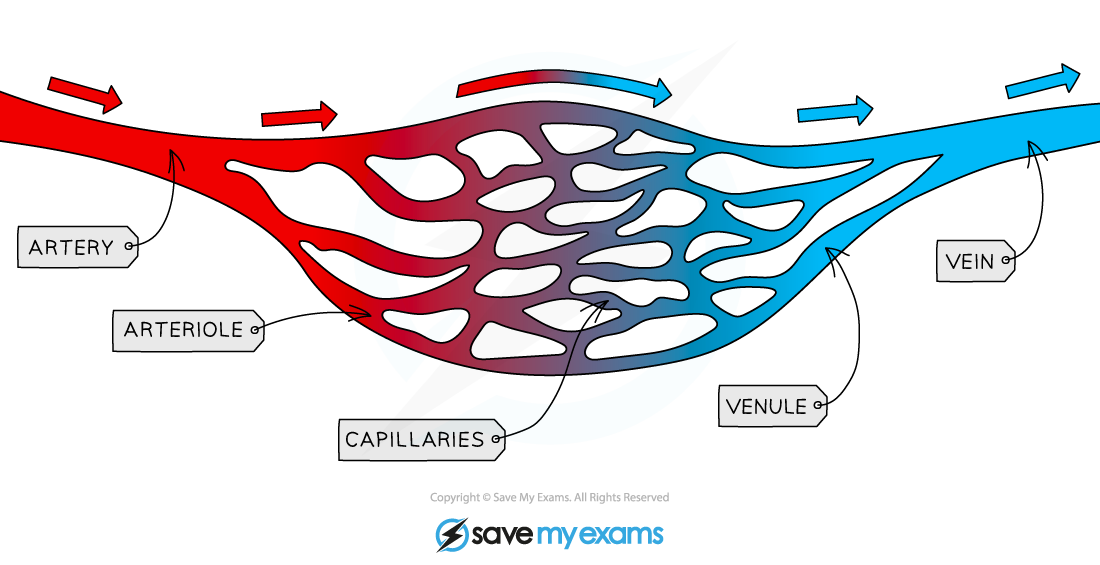
- Capillaries do not only transport blood, but they are the sites of material exchange between the blood and body tissues.
- They allow the exchange of oxygen, glucose, carbon dioxide, nutrients, and waste products between blood plasma and the surrounding tissue cells.
- Their walls are made of a single layer of endothelial cells (flat cells that form the inner lining of blood vessels). This feature provides an extremely short diffusion distance, allowing substances to move into and out of tissues rapidly.
- Capillaries form vast, intricate networks through every tissue in the body, providing a massive total surface area for efficient diffusion.
- The lumen is so tiny that red blood cells must squeeze through in a single-file line. This slows down the blood flow for maximum time for gas and nutrient exchange to occur.
Main blood vessels of the body.
To and from the heart
- Vena cava: Main vein that collects deoxygenated blood from the body organs and drops it into the right atrium.
- Aorta: Largest artery in the body. Leaves the left ventricle to deliver high-pressure, oxygenated blood to the rest of the circulatory system.
To and from the lungs
- Pulmonary artery: Carries deoxygenated blood away from the right ventricle to the lungs.
- Pulmonary vein: Returns oxygenated blood from the lungs back to the left atrium.
To and from the kidney
- Renal artery: Branches off the aorta to bring oxygenated blood containing metabolic wastes (e.g. urea) into the kidneys for filtration.
- Renal vein: Carries filtered, deoxygenated blood away from the kidneys and joins the vena cava.
To and from the liver
- Hepatic artery: Supplies oxygenated blood directly from the aorta to the liver tissue to fulfill its high oxygen demands.
- Hepatic vein: Carries deoxygenated blood from the liver back to vena cava.
- Hepatic portal vein: An important vessel. It carries nutrient-ricj blood directly from the digestive tract (stomach and intestines) straight to the liver.
- This allows the liver to sort, store, or detoxify absorbed nutrients before they are distributed to the rest of the body.