Gas Exchange
Diagram
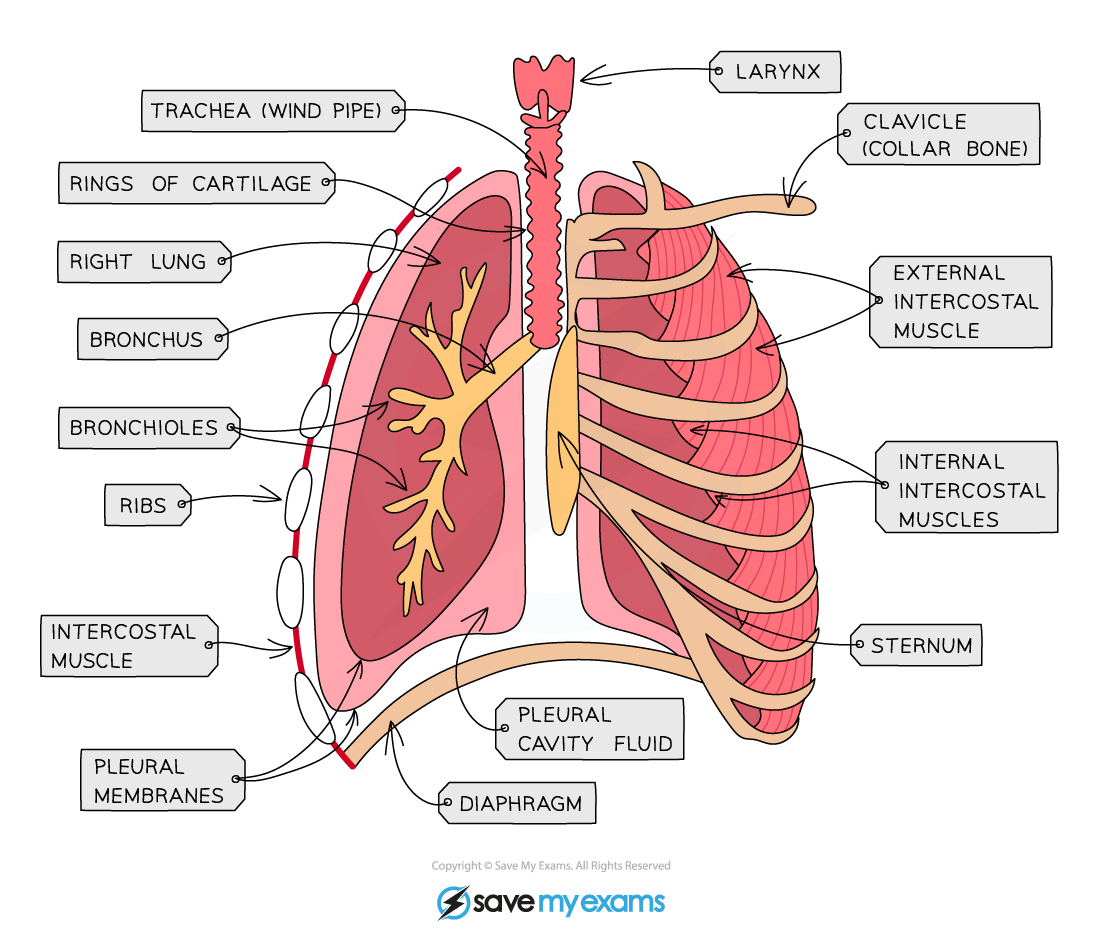
Parts of the respiratory system
- Larynx: Located at the top of the trachea. Contains vocal cords used to produce sound.
- Trachea: Main airway leading from throat to lungs.
- Function of cartilage: Trachea is lined with C-shaped rings of cartilage. These rings keep the trachea open at all times, preventing it from collapsing inward whent eh pressure drops during inhalation.
- Bronchi: Trachea splits into 2 tubes, the left and right bronchi, each leading into a lung.
- Bronchioles: Smaller, highly branched tubes branching off the bronchi. Distributes air throughout all parts of the lungs.
- Alveoli: Tiny air sacs at the end of the bronchioles where gas exchange actually takes place. Surrounded by a network of associated capillaries.
- Diaphragm: A dome-shapped sheet of muscle separating the thorax (chest cavity) from the abdomen.
Features of gas exchange surfaces
- Large surface area: Millions of alveoli collectively create a massive surface area, allowing large amounts of gas to diffuse simultaneously.
- Thin surface: The walls of both the alveoli and the capillaries are only one cell thick. This ensures a very short diffusion distance for oxygen and carbon dioxide.
- Good blood suppply: A dense network of capillaries constantly moves blood past the alveoli. This brings carbon dioxide-rich blood to the lungs and carries oxygenated blood away, maintaing a steep concentration gradient.
- Good ventilation with air: Breathing continuously replaces old, low-oxygen air with fresh, high-oxygen air, preserving the steep concentration gradient required for rapid diffusion.
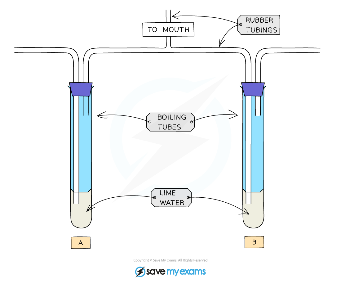
Investigating the differences (limewater test)
- To demonstrate that expired air contains significantly more carbon dioxide than inspired air, you can set up a simple boiling tube apparatus containing limewater.
- Carbon dioxide turns clear limewater milky or cloudy.
- Air is drawn through one flask when you inhale, and your exhaled breath is bubbled through a second flask when you exhale.
- The result is that the limewater in the exhalation tube turns cloudy almost instantly, whereas the inhalation tube remains clear for a long time, proving that expired air has a much higher concentration of carbon dioxide.
Inhalation vs. exhalation actions
| Feature/action | Inspiration (inhalattion) | Expiration (exhalation) |
|---|---|---|
| External intercostal muscles | Contracts (raises ribs upwards and outwards) | Relaxes |
| Internal intercostal muscles | Relaxes | Contracts (lowers ribs downwards and inwards) |
| Diaphragm action | Contracts and flattens | Relaxes and bulges up |
| Thorax volume | Increases | Decreases |
| Thorax pressure | Decreases (below atmospheric pressure) | Increases (above atmospheric pressure) |
| Air movement | Rushes into the lungs | Forced out the lungs |
Composition of inspired vs. expired air
| Gas | Inspired air (%) | Expired air (%) | Reason |
|---|---|---|---|
| Oxygen | 21 | 16 | Absorbed by diffusion into the blood at the alveoli to be used by cells for aerobic respiration |
| Carbon dioxide | 0.04 | 4 | Produced as a waste product of aerobic cellular respiration, carried by blood to lungs, and diffused out |
| Water vapour | Variable | Saturated (high) | Water evaporates from the moist lining of the gas exchange surfaces into the air sacs |
Effects of physical activity on breathing
- When you exercise, your muscles contract harder each time. This triggers a specific physiological loop.
The feedback loop
- Physical activity begins. Musccle cells increase their rate of aerobic respiration to produce more ATP for muscle contraction.
- Carbon dioxide accumulates. As a waste product of this respiration, the carbon dioxide concentration in the blood increases.
- Brain detects deoxygenated blood. The blood passes throught the brain, where specialized receptors detect this high carbon dioxide level.
- Signal is sent to the diaphragm and intercostal muscles.
- The response is an increase rate (more breaths per minute) and a greater depth (larger volume per breath of breathing.
- The purpose of the rapid gas exchange accelerates the removal of toxic carbon dioxide from the blood and supplies the extra oxygen needed by the muscles.
Protection of the breathing system
- The air we breathe contains dust, smoke particles, and pathogens. The respiratory tract uses a two-part defense mechanism to keep the lungs clean.
- Goblet cells are specialized cells scattered along the lining of the trachea and bronchi. They synthesize and secrete sticky mucus.
- Mucus traps dust, dirt, and pathogens before they can reach the alveoli.
- Ciliated cells are cells that have tiny, hair-like extensions called cilia.
- The cilia beat in a synchronized, wave-like motion, pushing the trapped mucus up and out of the airway toward the larynx/throat, where it can be safely swallowed or coughed out.