The Heart
Diagram
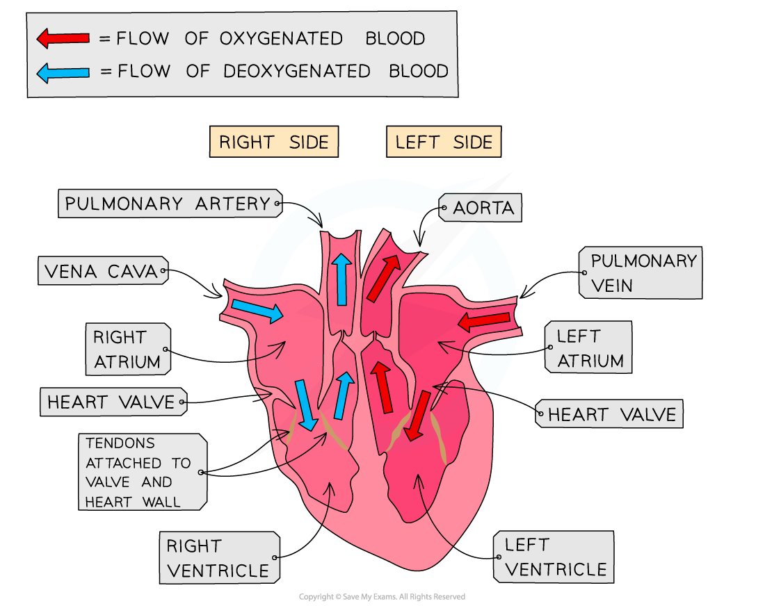
Structures of the heart
- Atria (sg. atrium): The two upper chambers. Thin walls and receive blood entering the heart from the veins.
- Ventricles: Two lower chambers. They have thick muscular walls and pump blood out of the heart into the arteries
- Septum: A solid wall of muscle that separates the left and right sides of the leart.
- It prevents oxygenated blood (left side) from mixing with deoxygenated blood (right side), ensuring the body receives the maximum possible concentration of oxygen.
- Valves: Flaps of tissue that keep blood flowing in one direction.
- Atrioventricular (AV) valves: Situated between the atria and ventricles. They stop blood from flowing back into the atria when the ventricles contract.
- Semilunar (SL) valves: Found at the entrances to the pulmonary artery and aorta. They prevent blood from flowing back into the ventricles when the heart relaxes.
- Coronary arteries: A network of blood vessels running across the outside of the heart. They branch off the aorta to supply the heart muscle tissue itself with its own constant supply of oxygenated blood and glucose.
- Blood is pumped AWAY FROM the heart by the ARTERIES.
- Blood is returned TO the heart by the VEINS.
Relative thickness of the ventricles and atria
| Comparison | Thickness difference | Explanation |
|---|---|---|
| Atria vs. Ventricles | Ventricles are much thicker than atria. | Atria only need to pump blood a very short distance (just down into the ventricles). Ventricles must pump blood out of the entire heart to the lungs or body. |
| Left vs. right ventricle | Left ventricle wall is significantly thicker than the right ventricle wall. | The right ventricle only pumps blood to the lungs (a short distance, requiring low pressure to protect delicate tissues). The left ventricle must pump blood to the entire rest of the body, overcoming high resitance and requiring much higher pressure. |
Monitoring heart activity
- The activity of the heart may be monitored by:
- ECG (electrocardiogram): Traces the electrical impulses passing through the heart muscle during the cardiac cycle.
- Pulse rate: Measuring the expansion and recoil of an artery (commonly at the wrist or neck) per minute as the left ventricle pumps blood.
- Listening to sounds of valves closing: Listening directly to the lub-dub sounds of the internal heart valves closing and opening
Effect of exercise
- When you exercise, your muscles contract much more frequently and powerfully.
- Active muscles require a significantly higher amount of energy, which they release via aerobic respiration.
- To sustain this, the muscles need an increased supply of oxygen and glucose. They also produce carbon dioxide waste much faster, which must be removed.
- The brain detects the increase of carbon dioxide in the blood and signals the heart to beat faster and contract harder (increasing stroke volume). This speeds up blood circulation to fulfill the metabolic demands of the working tissues.
Coronary heart disease
- Coronary heart disease is caused by blockage of the coronary arteries that supply the heart with glucose and oxygen.
Risk factors
- Diet: Consuming high amounts of saturated fats and cholesterol increases plaque buildup.
- Lack of exercise: Weakens the heart muscle and contributes to high blood pressure and obesity.
- Smoking: Nictonie constricts blood vessels and damages their linings, making plaque buildup more likely. Carbon monoxide reduces the blood’s oxygen-carrying capacity.
- Stress: Releases hormones that chronically elevate blood pressure, putting strain on the arteries.
- Genetic predeposition: A family history of cardiovascular disease can mean naturally higher blood pressure or cholesterol levels.
- Age: The risk increases naturally as blood vessels lose their elasticity over time.
- Sex: Males statistically face a higher risk than pre-menopausal females due to the protective effects of oestrogen.
Prevention (roles of diet and exercise)
- Diet adjustments: Replacing saturated fats with unsaturated fats, reducing salt intake (lowers blood pressure), and increasing dietary fiber helps lower blood cholesterol levels and reduces the rate of plaque buildup.
- Regular exercise: Strengthens the heart muscle, increases cardian efficiency (lower resting heart rate), lowers systemic blood pressure, and helps metabolize excess lipids/cholesterol.
Mechanics of a heartbeat
- Contraction of atria: The muscles of the left and right atria contract simutaneously. This squeezes blood through the open atrioventricular valves into the ventricles. The semilunar valves remain shut.
- Contraction of ventricles: The ventricles contract from the bottom up. This rise in pressure forces the atrioventricular valves shut to prevent backflow to the atria and also to create the first “lub” sound of the heartbeat. The pressure forces the semilunar valves open, shooting blood into the pulmonary artery and the aorta.
- Heart relaxes: The heart muscles relax. To prevent the high-pressure blood in the arteries from slipping back down into the ventricles, the semilunar valves snap shut to create the second “dub” sound. Blood from the vena cava and pulmonary vein trickles back into the passive atria, and the cycle repeats.